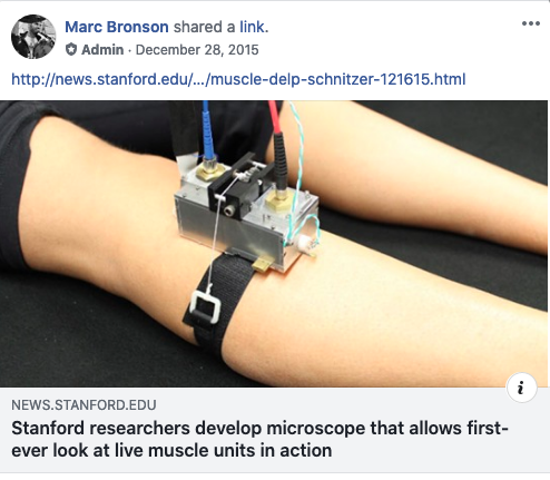
Stanford researchers develop microscope that allows first-ever look at live muscle units in action The wearable microscope allows precise measurements of sarcomere lengths and contractile dynamics in alert humans. Unlike traditional endoscopy, which is well suited for imaging in natural body cavities or those created intra-operatively, our instrument’s microendoscope works well in solid tissue and is propelled into the body in a fraction of a second. The microscope’s mobility (Figure 1A, inset), versatility (Figure 2), and minimally invasive capabilities allow new types of observations (Figures 3, 4, and 5) for medical research and clinical imaging of patients with movement disorders. Beyond muscle, the microscope should allow in vivo imaging of multiple tissues previously examined in vitro using SHG contrast, including skin, cartilage, tendon, bone, and tumors (Campagnola, 2011, Chen et al., 2012). The technological advances reported here should also apply to other forms of nonlinear optical contrast, such as coherent anti-Stokes Raman scattering, two-photon excited fluorescence, third-harmonic generation, and stimulated Raman scattering, which all show promise for tissue inspection but have been nearly prohibitive to deploy clinically due to the challenges of miniaturization.
#science #chiropractor #chiropractic #research #education #evidence based #patient centered #interprofessional #collaborative #rehabilitation #public health #spinal health #musculoskeletal health #ethics #pain #function #disability #QOL #knowledgetranslation